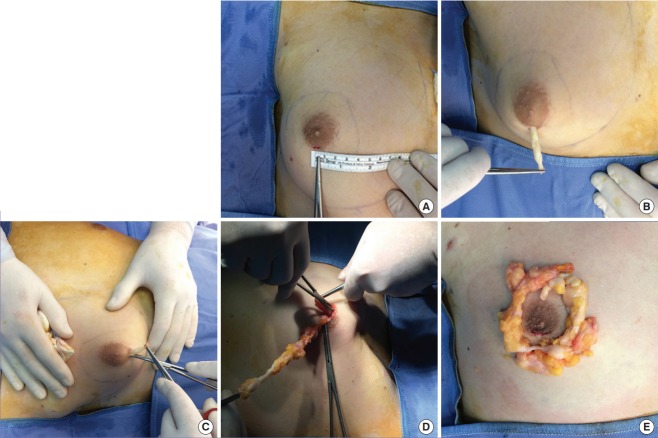
Fig. 1. Surgical technique.
(A) Surgery was performed under local anesthesia and a 3-mm skin incision was made along the inferior line of the areola at 6 o’clock. (B-D) By lifting the subcutaneous tissue under the nipple–areola flap, it was possible to access the glandular tissue with a 360° range under direct vision. Through the small incision, blunt dissection was performed in a clockwise and counterclockwise direction, describing two half-circles. The breast tissue was pulled out, held with a Kocher clamp, and the gland was transected along its length. (E) Two or more pieces were removed with forceps in a half-circle fashion.

