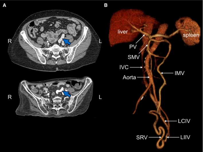
Figure 3.

Imaging of portosystemic shunt. A, CT scans. Blue arrow indicates dilated superior rectal vein, present from 6 months prior to first hospital admission (upper panel) to 8 years later (lower panel). Note dramatic weight loss. B, CT reconstruction of vascular system. Note shunt of superior rectal vein (SRV) to the left internal iliac vein (LIIV). The shunt produces early opacification of the left common iliac vein (LCIV), but not the right common iliac vein. Other abbreviations: IVC, inferior vena cava; IMV, inferior mesenteric vein, PV, portal vein; SMV, superior mesenteric vein
