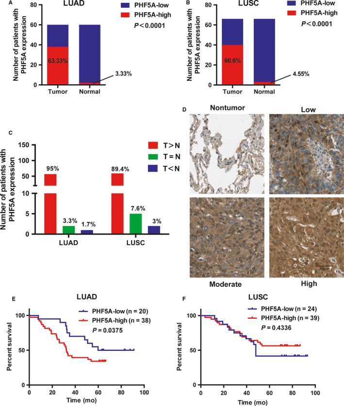
Figure 2.

The expression of PHF5A is significantly upregulated in both lung adenocarcinoma (LUAD) and lung squamous cell carcinoma (LUSC) patient samples. A and B, The percentage of PHF5A‐low and PHF5A‐high staining in tumor and paired normal tissues in LUAD (A, n = 60) and LUSC (B, n = 66) patients. C, The number and percentage of patients with higher, equal or lower PHF5A staining in LUAD and LUSC tumor tissues compared with paired normal tissues. T: tumor tissue; N: paired normal tissue. D, Representative IHC images of PHF5A staining in NSCLC tumor tissues and paired normal tissues. E and F, Kaplan‐Meier plots based on the expression level of PHF5A measured by IHC in 58 LUAD (E) and 63 LUSC (F) patients
