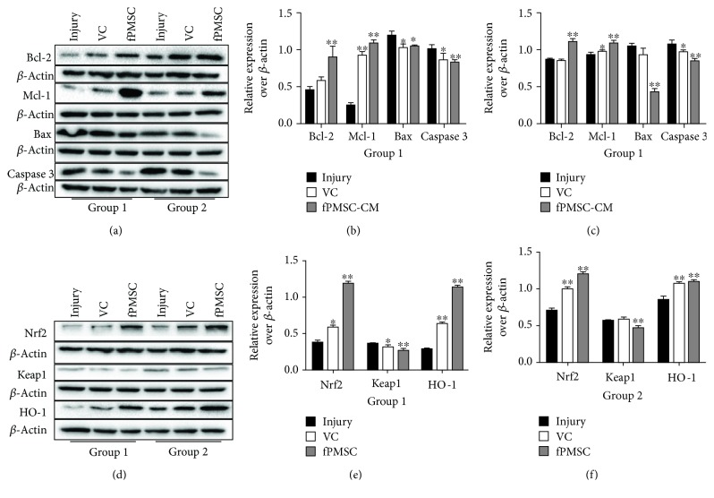
Figure 6.
Immunoblotting analysis of cell apoptosis and Nrf2-Keap1-ARE signaling proteins. The A549 cells were injured with 600 μmol/L H2O2 prior to being treated with 100 μmol/L VC or cocultured with hfPMSCs (3.0 × 10⁶/well) (group 1), or A549 cells (3.0 × 10⁶/well) were added with 100 μmol/L VC or cocultured with hfPMSCs (3.0 × 10⁶/well) for 24 h, then the medium of the bottom was exchanged with 600 μmol/L H2O2 for 24 h (group 2). (a) Immunoblotting assay determined Bax, Bcl-2, caspase 3, and Mcl-1 expression. (b, c) Semiquantitative analysis for proteins of interest by densitometry assay in (a) both group 1 (b) and group 2 (c), respectively. (d) Immunoblotting assay determined Nrf2, Keap1, and HO-1 expression. (e, f) Semiquantitative analysis for proteins of interest by densitometry assay in (d) both group 1 (e) and group 2 (f), respectively. Data represented the mean ± SD from three independent triplicated experiments (N = 9, t-test). Compared to the injury group, ∗ and ∗∗ represent p < 0.05 and p < 0.01, respectively.

