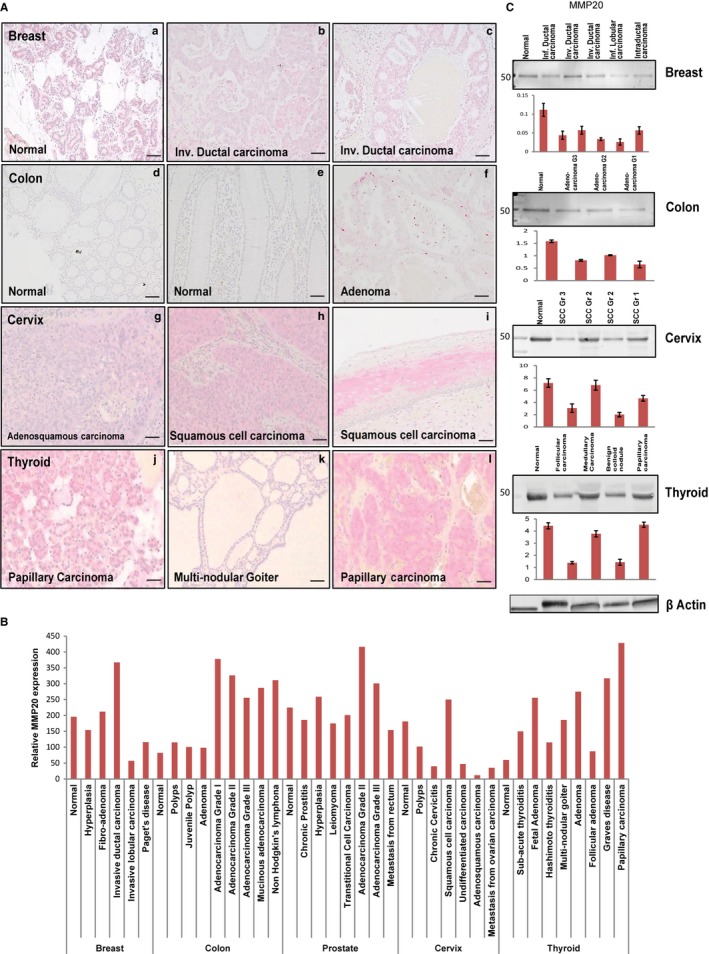
Figure 1.

MMP20 is expressed in the normal and neoplastic breast, colon, prostate, cervix, and thyroid tissue sections. A, Immunohistochemistry (IHC) on tissue microarrays (TMA) showing positive immunoreactivity for MMP‐20 in normal and invasive ductal carcinoma of breast tissue (a‐c); normal and adenoma colon tissue (d‐f); normal and squamous cell carcinoma of cervix tissue (g‐i); and, normal and papillary carcinoma of thyroid tissue (j‐l). B, Histogram for quantitative IHC of TMA shows differential expression of MMP‐20 in the normal and different grades of cancer in the breast, colon, prostate, cervix, and thyroid. C, Western blot (WB) and quantitative (fold change) histogram shows significantly high MMP‐20 protein levels in normal and cancer whole tissue lysates from the breast, colon, prostate, cervix, and thyroid tissues. β‐actin was used as the normalization control. Values are mean ± SE, n = 3. Data are representative of three independent experiments. Scale bar, 100 μm
