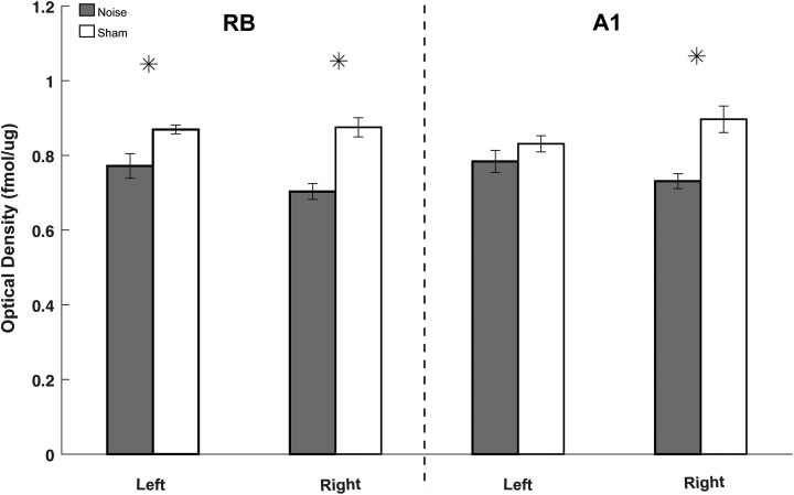
Figure 4.
Noise decreases mAChR expression in A1 and rostral belt (RB). Bar graph comparing quantified [3H]scopolamine binding pooled from all cortical subregions (supragranular, granular, and infragranular) from each respective anatomic location (RB and A1). Note that noise-exposed animals (gray) showed significantly decreased [3H]scopolamine binding in both hemispheres of RB (larger decreases contralateral to noise) and in the right hemisphere of A1 as compared to sham controls (white). Overall, [3H]scopolamine binding decreases similarly in both A1 and RB (* P < .05 as compared to respective sham control; standard deviation). mAChR indicates muscarinic acetylcholine receptor.

