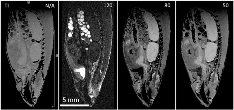
FIGURE 10.

Sagittal slice from a series of 3D IR FLASH IR images of Gd doped (1mM GdDTPA‐BMA, Omniscan) FF zebrafish. Each image with isotropic resolution of 50 μm/pxl, TE 2, TR 350, EncMtx 333 × 148 × 74, Mtx 400 × 176 × 88, FOV 20 × 8.8 × 4.4, tt 4 h 15 min. A, No inversion; B, TI 120; C, TI 80; D, TI 50. Judicious selection of inversion time affords contrast enhancement of organs of interest
