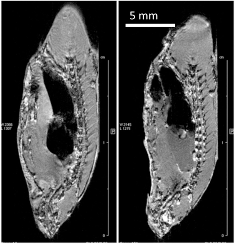
FIGURE 9.
Sagittal slices through 3D FLASH images of fresh (unfixed) zebrafish: VC20, TR 50, TE 2.6, EncMtx 512 × 160 × 100, Mtx 512 × 160 × 100, FOV 32 × 10 × 6.4, Resol 64 × 64 × 64. Left: immediately after sacrifice, NAV 32, tt 7 h 6 min. Right: after 48 h in the scanner at 4°C, NAV 22, 4 h 53 min. The specimen changes shape over time due to migration of liquid into the swim bladder

