
Fig. 2. Workflow.
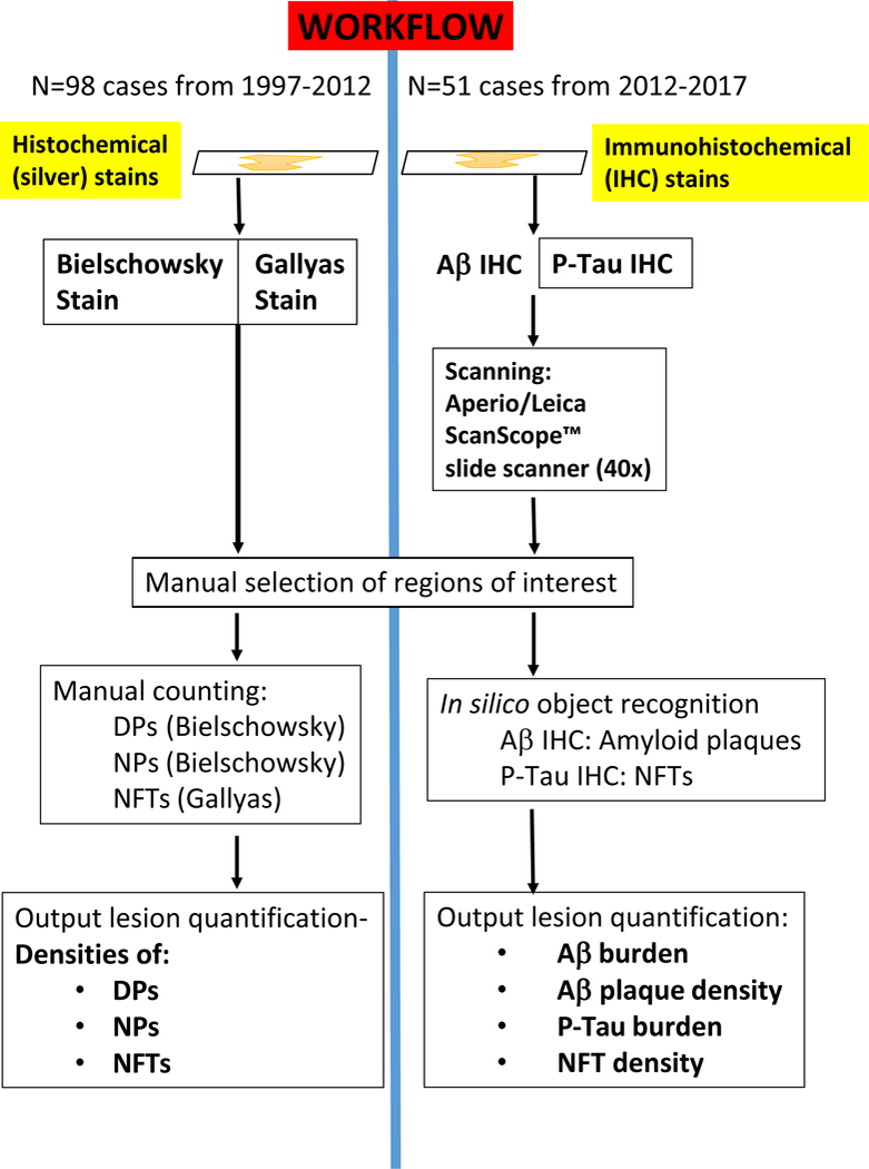
Schematic to depict the study design of the present study that included different UK-ADC cohorts and methods applied. Notably, different stains were used and the neuropathologic lesions were detected and counted separately. For the 1997–2012 cohort, histochemical stains were used with silver impregnation. Bielschowsky stains were used for diffuse amyloid plaques (DPs) and neuritic amyloid plaques (NPs), whereas the Gallyas silver impregnation technique was used for NFTs; for these cases, the output comprised manually tabulated density counts for DPs, NPs, and NFTs. For the 2012–2017 cohort, immunohistochemical (IHC) stains were used -- Aβ and P-Tau IHC – and slides scanned using a digital slide scanner, followed by in silico object recognition, with output including Aβ burden, Aβ plaque density, P-Tau burden, and P-Tau NFT density.