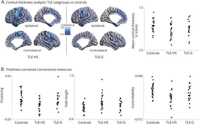
Figure 4. Robustness of white matter connectome measures with respect to cortical thickness.
(A) Surface-wide group analysis of cortical thickness between temporal lobe epilepsy with hippocampal sclerosis (TLE-HS) and temporal lobe epilepsy with gliosis (TLE-G) compared to healthy controls. Maps show pFDR < 0.05 corrected findings (black outlines) superimposed on Cohen d effect size maps (thresholded at d > 0.2, semitransparent). Although effect sizes were larger in TLE-HS, there was no significant difference between patient subgroups after the correction for multiple comparisons. Whole-brain mean cortical thickness findings are displayed for the 3 cohorts. (B) Thickness-corrected measures of clustering coefficient, path length, and controllability in the 3 groups, highlighting robustness of the marked effect of TLE-HS on white matter connectome organization.

