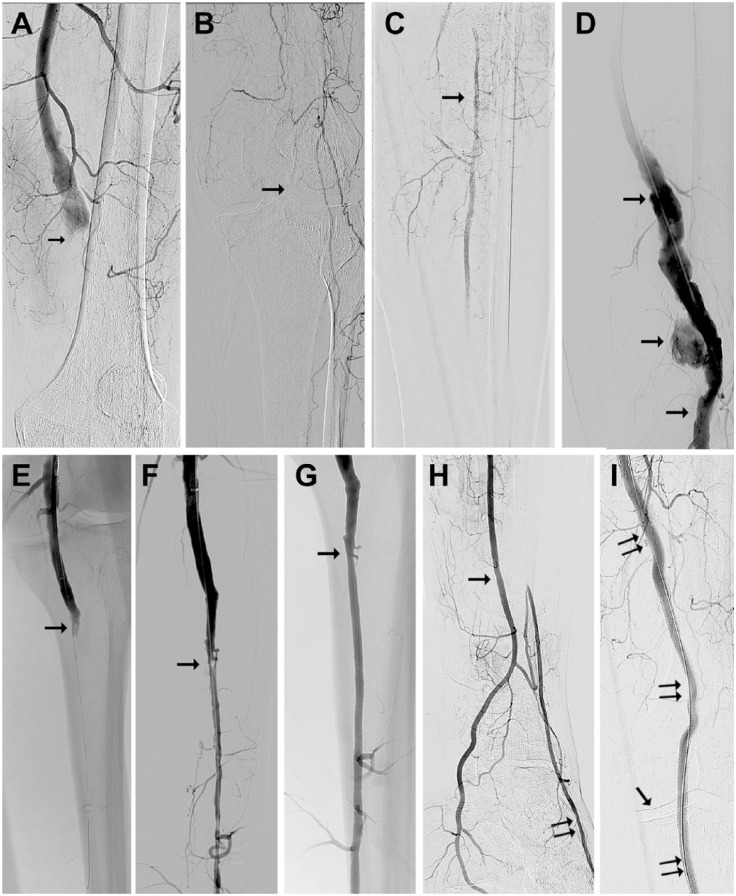
Figure 1.
(A) Digital subtraction angiography from a 56-year-old man with acute limb ischemia type IIb owing to popliteal aneurysm thrombotic occlusion (arrow). (B) The knee joint is depicted by the arrow. (C) Only one fragment (arrow) of the calf vessel is filled with contrast via collaterals. (D) After mechanical debulking of the popliteal artery with recanalized lumen (arrows). (E) The popliteal artery was recanalized to its periphery (arrow) with the Rotarex catheter. (F) The popliteal artery debulking opened the way for successful dilation of the peroneal artery with residual stenosis (arrow). (G) The stenosed area after dilation and stenting (arrow). (H) The peroneal artery (arrow) supplies the plantar vessels directly and the dorsal pedis artery via collaterals (double arrow). (I) The popliteal aneurysm was excluded using a Viabahn stent-graft (double arrows); the single arrow indicates the knee joint.

