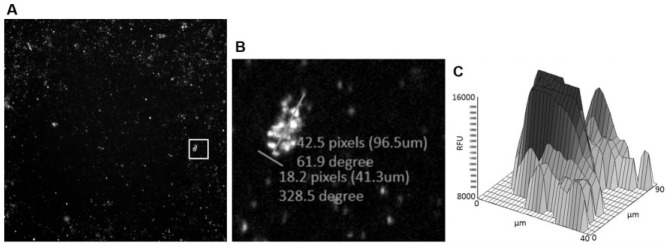
Figure 3.
(A) Operetta image (4× objective) of 384-well plate containing C6/36 (wAlbB) cells infected with Wolbachia. Primary antibody against wBmPAL with Alexa Fluor 680 secondary antibody. White box denotes Wolbachia clump. (B) Zoomed view into area denoted by white box in (A) showing Wolbachia clump with measuring lines. (C) Corresponding acumen 2D fluorescence intensity plot of a Wolbachia clump.

