
Fig. 5.
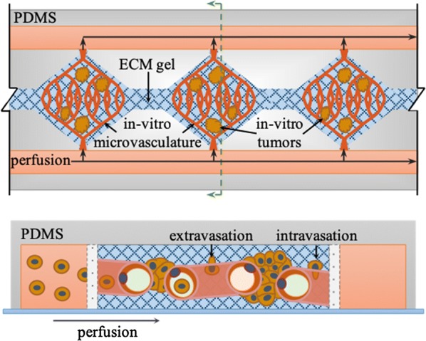
On-chip biomimetic model to study metastatic lung cancer. Tumor microvasculature-on-a-chip. Top-view (top) and cross section (bottom) of a multicompartment microfluidic chip for the development of perfusable microvascular networks and microtumors. Diamond-like chambers support the growth of microvascular networks emended in extracellular matrix (ECM) gels, while flanking side channels are used to perfused nutrients and drugs. Perfusable microvascular networks are formed by co-culturing microvascular endothelial cells with lung fibroblast and vascular smooth muscle cells in the ECM gel. Lung cancer cells can be co-injected before ECM gelification to grow microtumors. Alternatively, lung cancer cells can be perfused via flanking channels to study metastatic colonization (adapted from Sobrino et al. [190])