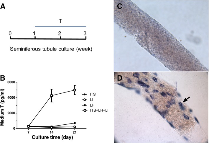
Fig. 1.
An in vitro culture system for differentiating stem Leydig cells on the surface of seminiferous tubule into Leydig cell lineage. Panel a-b: The testosterone production by seminiferous tubules in the BM contained ITS 5 mM, LH 5 ng/ml, LI 5 mM, or ITS 5 mM + LH 5 ng/ml + LI 5 mM (the LIM). Mean ± SEM, n = 4–8. Panel c-d: Seminiferous tubules were stained with HSD3B1 and Leydig cells were indicated by black arrows. Panel: (c) Seminiferous tubule after 21 days of culture in the BM (no Leydig cells were observed); (d) Leydig cells were observed with HSD3B1 staining (cells indicated by black arrow on the surface of a seminiferous tubule) after 21 days of culture in the LIM

