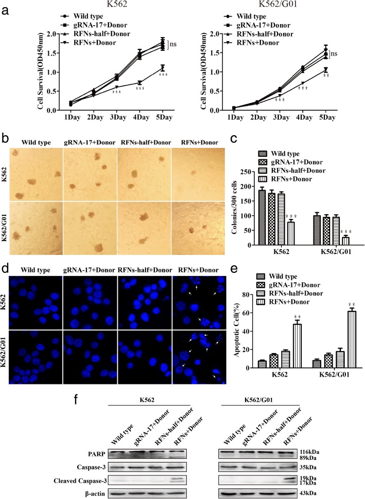
Fig. 4.
RFNs suppress viability and induce apoptosis of imatinib sensitive and resistant cells. Cells were transfected with gRNA-17 plus donor, RFNs-half plus donor, RFNs plus donor, respectively. a Evaluation of cell proliferative capacity by CCK-8 assay. b, c The capacity of colony formation was assessed by colony-forming assay. d Cell nucleuses were stained by DAPI to detect the morphologic changes caused by apoptosis. The white arrows pointed out the typical apoptotic cells. e Satistical analysis result of apoptotic cells tested by flow cytometry. f The activation of apoptotic pathway was investigated by western blot. The results are presented as the means ± SD. One-way ANOVA analysis was used to compare treated groups with control group, p < 0.01 (**) and p < 0.001 (***)

