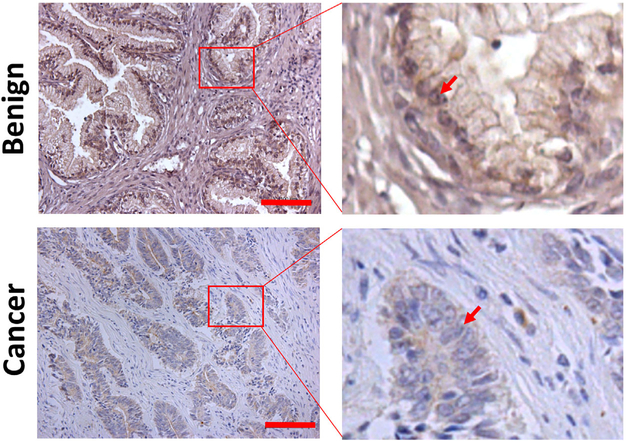
FIGURE 1.
Immunostaining of ELL2 in clinical prostate cancer specimens. The red bars in the left panel of the images indicate 100 μm. The red arrows indicate the nuclei of luminal epithelial cells in the benign prostate tissue or the nuclei of cancer cells. [Color figure can be viewed at wileyonlinelibrary.com]

