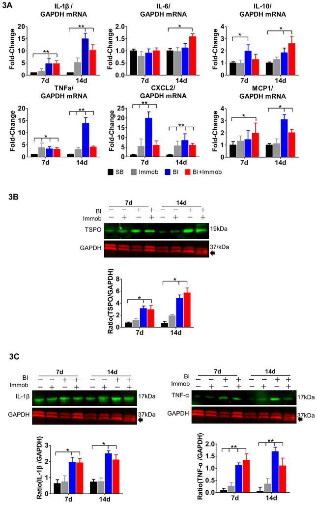
Fig. 3. Pro-inflammatory cytokines and chemokines expression in spinal cord after distant body burn injury.
(A) Quantitative PCR mRNA expression of cytokines (IL-1β, IL-6, IL-10 and TNFα) and chemokines (CXCL2 and MCP1) were measured and normalized to GAPDH. The mRNA expression of pro-inflammatory and chemokine molecules, IL-1β, TNF-α, and CXCL2, MCP-1, was sharply increased at day 7 and/or 14 post-injury in BI or BI+Immob groups relative to SB. The mRNA expression of TNF-α and CXCL2 was significantly increased in Immob alone group compared to SB. (B) Immunoblots of lumbar spinal cord (L3-L4 segments) isolated from SB, Immob, BI and BI+Immob mice at day 7 and 14 post-injury probed for TSPO and normalized to GAPDH as loading control and fold change of the immunoblot was determined using Image J to measure band intensities of target protein to GAPDH. The expression of TSPO, a marker of microglial activation, was significantly upregulated in BI and BI+Immob at both 7 and 14 days post perturbation. (C) Immunoblot of lumbar spinal cord (L3-L4 segment) protein isolated from SB, Immob, BI and BI+Immob mice at day 7 and 14 post-injury probed for IL-1β, TNF-α and normalized to GAPDH using Image J to measure band intensities of target proteins. The immunoblots confirm microglia activation and pro-inflammatory cytokine (IL-1β, TNF-α) release.to GAPDH (loading control). Data are means ± SE; SB: n = 6, Immob-7d: n=5, BI-7d: n = 6, BI+Immob-7d: n = 6, Immob-14d: n=5, BI-14d: n = 5, BI+Immob-14d: n = 6. *p < 0.05 for BI or BI+Immob vs. SB, **p < 0.001 for BI or BI+Immob vs. SB.

