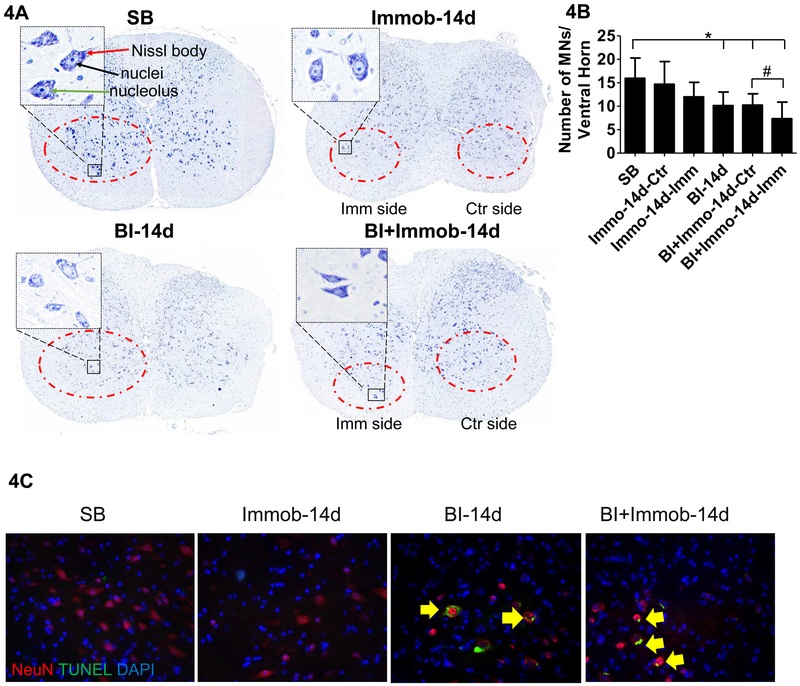
Fig. 4. Burn injury-induced apoptosis with decrease ventral horn motor neurons.
(A) Nissl staining of spinal cord dorsal and ventral horn. Imm side refers to immobilized side and Ctr refers to non-immobilized side of same animal. Compared to SB which show many Nissl stained bodies in the ventral horn, the BI group and BI+Immob groups showed decreased Nissl staining at day 14. As same as SB group, Immob group motor neurons showed regular shape, clear nucleus and Nissl bodies in the cytoplasm. The ventral horn image is magnified (400X) on the left dorsum of figure. The magnified images show motor neurons in SB, Immob, BI and BI+Immob groups at day14 after perturbations. The Nissl body is indicated by red line, the nuclei by black line and, the nucleolus by green line. The cytoplasm with a clearly visible nuclei and nucleolus is seen in SB. In contrast, fewer morphologically normal neurons were visible in BI and BI+Immob group, especially in BI+Immob group Imm side. Motor neurons cells were swollen with shrunken cytoplasm or vacuolation; pyknosis was also observed in BI-14d and BI+Immob-14d groups. (B) Ventral horn motor neurons (diameter ≥25 μm) counts in spinal cord, indicate that morphologically normal motor neurons decreased after BI and BI with immobilization. Data are means ± SE; SB: n = 12, Immob-14d: n = 10, BI-14d: n = 10, BI+Immob-14d: n = 12. *p < 0.05 for BI or BI+Immob vs. SB, #p < 0.05 for BI+Immob Imm side vs. BI+Immob Ctr side. (C) Double immunofluorescence staining and TUNEL staining for ventral horn motor neuron in spinal cord. The images show that BI and BI+Immob increased apoptotic cells in ventral horn motor neurons. Magnification 20×, red: motor neuron, green: TUNEL signal, blue: nucleus, yellow arrow indicates apoptotic motor neurons.

