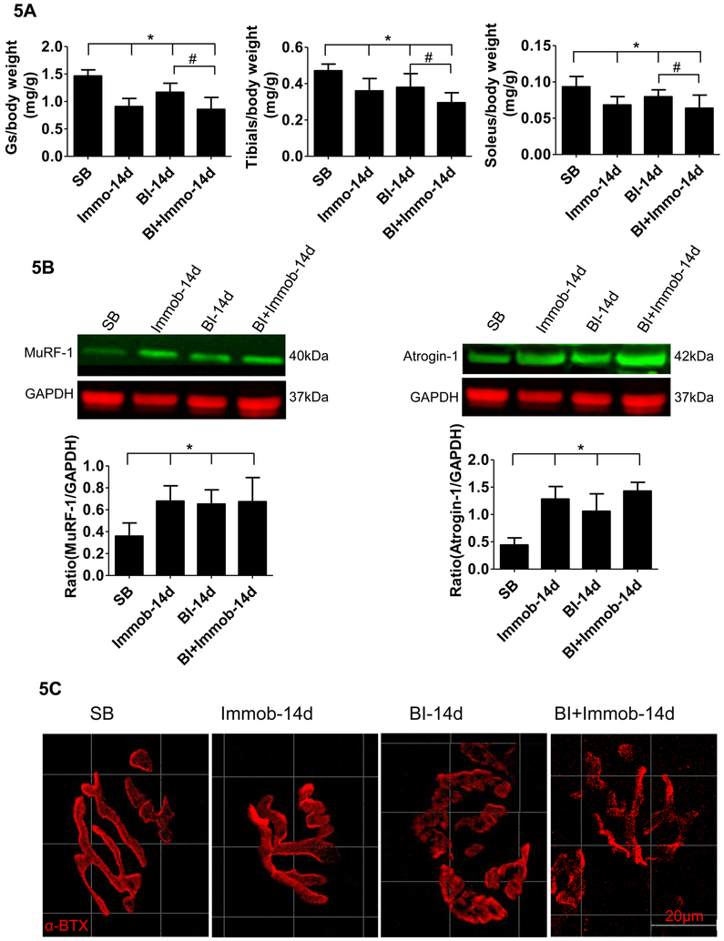
Fig. 5. Burn injury induced muscle wasting and neuromuscular junction synapse disintegration.
(A) The changes in muscle weight of SB, Immob, BI and BI+Immob group mice at day 14 post-injury. Dry muscle mass (gastrocnemius (Gs), soleus and tibialis anterior muscle) is expressed as a ratio to body weight at day preborn Day. The body weight ad day 0 was used in view of body weight loss induced by BI at day 14. Those histograms show that gastrocnemius, tibialis anterior and soleus muscles masses were decreased ~19- 38% at 14 days in Immob, BI, and BI+Immob group. Data are means ± SE; n = 5–8 mice/group. *p < 0.05 for Immob, BI or BI+ Immob vs. SB, #p<0.05 for BI+Immob vs.BI. (B) Immunoblots of muscle specific ubiquitin ligases, MuRF-1 and atrogin-1 expression in gastrocnemius muscle, and quantitation of MuRF-1 and atrogin-1 expression normalized to GAPDH is shown. Those histograms show that MuRF-1 and atrogin-1 expression was significantly increased in the Immob, BI and BI+Immob groups. Data are means ± SE; n = 4 mice/group. *p < 0.05 for Immob, BI or BI+ Immob vs. SB; (C) Gastrocnemius muscles were stained with α-BTX (red color) to detect acetylcholine receptors at the muscle endplates (synapse). The endplate in SB and Immob group appear compact and pretzel shaped. In BI and BI+Immob group the synapses were fragmented, more marked in the latter group, evidenced by the loss of typical pretzel shape. Scale bar = 20 microns.

