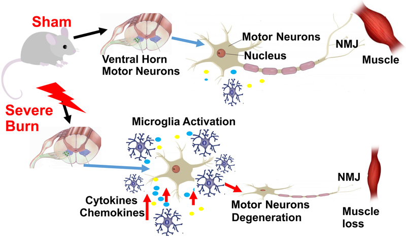
Fig. 6. Schematic diagram of motor neurons and microglia in spinal cord without or with burn injury.
Cartoon of motor neuron unit of SB (normal) (upper panel) and BI (lower panel) mice. The cartoon demonstrates that in the unburned state that there is minimal microglial activation in the spinal ventral horn and no changes in synapse or muscle mass. Severe BI induces microglia activation in spinal cord ventral horn, evidenced as inflammatory cytokine and chemokine release and is associated with degeneration of motor neurons that correlates to skeletal muscle mass loss.

