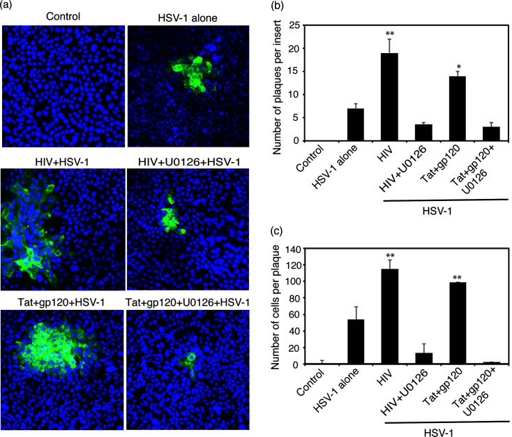
Fig. 4.
HIV-induced activation of MAPK/NF-κB and MMP-9 facilitated cell-to cell spread of HSV-1. (a) Polarized tonsil epithelial cells were exposed to cell-free HIV-1SF33 virions and tat+gp120 in combination for 5 days, with or without U0126. On day 5, epithelial cells were infected with HSV-1 from the basolateral surface and incubated for 3 days. Cells were fixed and then immunostained for HSV gD (green). Cell nuclei were stained blue. (b) HSV-1-infected plaques were counted on three inserts for each experiment. Results are presented as the average number of plaques or average number of infected cells per plaque. (c) HSV-1-infected cells were quantified in 10 random microscopic fields per plaque. Error bars indicate sem (n=3). *P<0.005, **P<0.001, all compared to the control group. Two independent experiments showed similar data.

