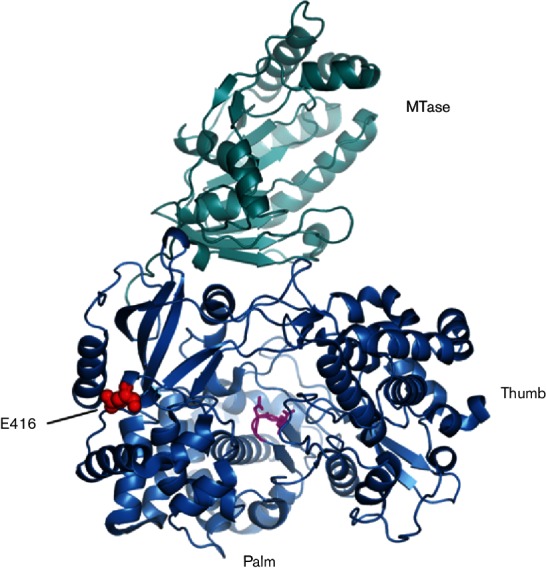
Fig. 4.

Structure of the NS5 protein with SLEV-E416K. The MTase domain (cyan) and polymerase domain (blue) are shown in ribbon form looking into the active site. Residue E416 is displayed as space-filling spheres (red), and the catalytic GDD sequence (motif C) is in stick representation (magenta).
