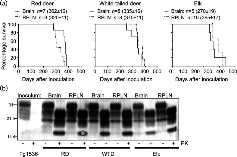
Fig. 2.
Prions from the CNS and lymphoreticular system of CWD-infected deer and elk are transmissible to CWD-susceptible transgenic mice. (a) Tg(DeerPrP) 1536 mice were intracerebrally inoculated with 1 % homogenates of brain and RPLN tissues from CWD-infected cervids. Kaplan–Meier survival curves are shown for each infected cohort. Mean incubation times (±SEM), where n indicates the number of infected and diseased animals, are also shown. Asterisks indicate statistical significance between the mean incubation periods of matched samples (*, P<0.05; **, P<0.005, Mantel–Cox). (b). All of the diseased Tg(DeerPrP) 1536 mouse brains were analysed by immunoblotting to confirm prion disease. Samples from two representative mice in each group are shown. The inoculum tissues are listed above the blots, and the inoculum species and PK treatments are listed below the blots.

