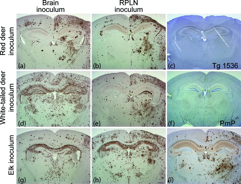
Fig. 3.
The PrPSc deposition patterns in mice varied with inoculum source. We prepared coronal sections through the hippocampus of Tg(DeerPrP) 1536 mice inoculated with: (a) red deer brain, (b) red deer RPLN, (c) nothing (uninoculated), (d) white-tailed deer brain, (e) white-tailed deer RPLN, (g) elk brain, (h) elk RPLN, 320 days p.i., (i) elk RPLN, 400 days p.i. We used prion knockout mice (PrnP−/−) as a negative control for IHC (f). We stained paraffin-embedded sections with anti-PrP antibody 18 days after formic acid treatment and antigen retrieval. Scale bar (h), 500 µm. All mice were inoculated in the right hemisphere.

