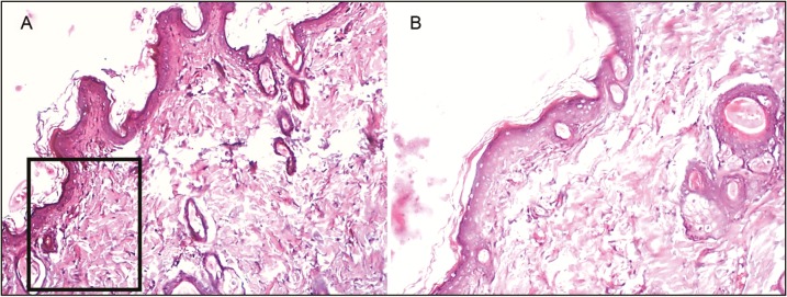
Figure 8.
Histopathological slides for the diabetic standard group. Figure (A) and (B) represents wound tissue specimen (wound bed) after complete healing of excision wound, stained with hematoxylin and eosin in the diabetic group (group VII: standard silver sulfadiazine) as viewed under light microscope 4× and 10×. The sections showed well-formed keratinized squamous epithelium and the underlying dermis showed loosely arranged collagen fibers

