Figure.

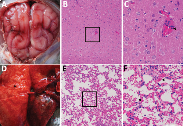
Dissected brain and lung of a dead fox, collected in 2017 in Shandong Province, eastern China, and histopathologic examination of samples using hematoxylin and eosin staining. A) Brain, showing congestion in the meninx. B) Histologic view of meninx, showing mild neuronal degeneration and inflammatory cell infiltration in vessels. Original magnification ×100. Box indicates area enlarged in panel C. C) A higher magnification view (original magnification ×400) of lesions in panel B, showing inflammatory cell infiltration in a vessel (arrow). D) Lung tissue, showing extensive congestion and hemorrhage. E) Histologic view of lung tissue, showing congestion, hemorrhage, or both, with many erythrocytes in the alveolar space. Original magnification ×100. Box indicates area enlarged in panel F. F) A higher magnification view (original magnification ×400) of tissue lesions in panel E, showing erythrocytes in the alveolar space (arrows).
