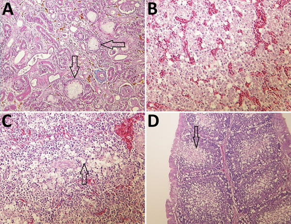
Figure 7.

Histologic appearance of lesions in tissues from chickens experimentally infected with a novel orthobunyavirus (Kedah fatal kidney syndrome virus) isolated from broiler chickens with severe kidney disease, Malaysia, 2014–2017. A) Kidney, showing urate deposits in the dilated Bowman’s capsule (arrows) and degeneration and dilatation of proximal convoluted tubules. Trichcrome stain; original magnification ×200. B) Liver, showing vacuolar degeneration of hepatocytes and disorganization of lobular structure. Hematoxylin and eosin (H&E) stain; original magnification ×400. C) Spleen, showing marked lymphoid depletion and lymphocyte necrosis in the Malpighian body and vacuolation of reticulocytes in the red pulp (arrow). H&E stain; original magnification ×400. D) Bursa Fabricii, showing atrophy of bursal follicles with marked lymphoid depletion in the medullary region (arrow). H&E stain; original magnification ×200.
