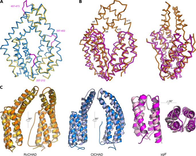
Figure S4. CHAD domains are formed by two 4-helix bundles related by an internal pseudo twofold symmetry.
(A) Structural superposition of the CtCHAD structure (in blue) with a second CtCHAD crystal form (PDB-ID 3E0S, in yellow; r.m.s.d. is ∼0.6 Å comparing 290 corresponding Cα atoms). Shown are Cα traces, with loop regions present in our structure and missing in PDB-ID 3E0S highlighted in magenta. (B) Structural superposition of RcCHAD (in orange) with the E. coli ygiF CHAD domain (PDB-ID 5A61, residues 205–428, in magenta) reveals the presence of protruding helices in RcCHAD forming the central pore (r.m.s.d. is ∼2.4 Å comparing 184 corresponding Cα atoms). (C) Structural superposition of the two 4-helix bundles in RcCHAD (residues: 7–166 versus 167–303), CtCHAD (residues: 207–373 versus 374–522), and ygiF (residues: 205–336 versus 337–428) shown as ribbon diagrams in two orientations.

