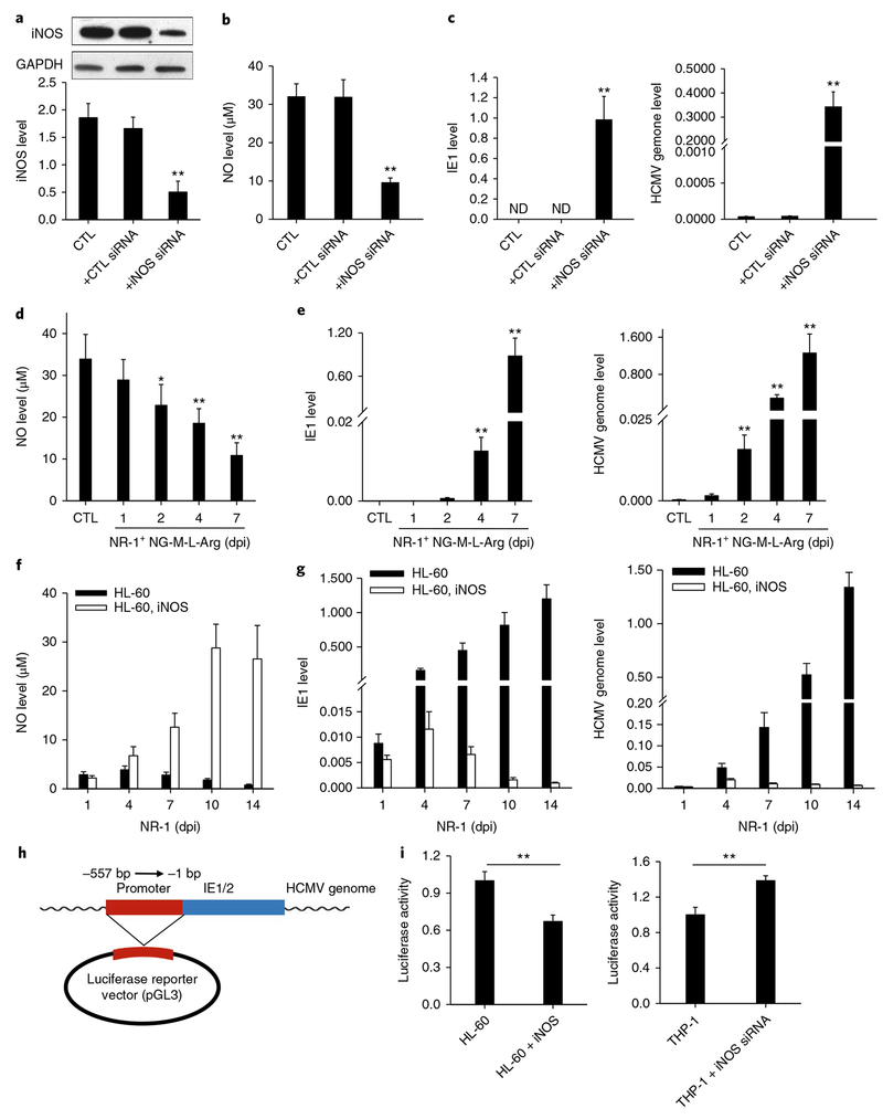
Fig. 4 |. Cellular iNOS/NO induced by STAT3 activity play a critical role in suppressing HCMV IE1 expression and viral replication in HPCs.
a,b, Levels of iNOS (a) and NO (b) in HPCs transfected with iNOS siRNA or scramble oligonucleotide. c, Increase of HCMV IE1 expression and genome replication in NR-1-infected HPCs after iNOS siRNA transfection. d, Depletion of cellular NO by NG-M-L-Arg. e, Increase of HCMV IE1 expression and genome replication in NR-1-infected HPCs after direct depletion of cellular NO by NG-M-L-Arg. f, Lentivirus-mediated iNOS overexpression in HL-60 cells increased intracellular NO level. g, Increase of NO level in HL-60 cells suppressed viral IE1 activity and genome replication. h,i, Inhibition of cellular NO on the activity of HCMV IE1 promoter. A luciferase reporter consisting of IE1/2 promoter region was constructed in pMIR-REPORT plasmid (h) and then transfected into HL-60 and THP-1 cells, respectively. HL-60 and THP-1 cells were treated with LV-iNOS or LV-iNOS siRNA to overexpress or knock down iNOS, respectively, prior to NR-1 infection at MOI of 2. Cellular luciferase activity (i) was assayed. Data are presented as the mean ± s.e.m. of three independent experiments. *P < 0.05, **P < 0.01 as determined by the two-tailed t-test (the P values are detailed in Supplementary Table 1). LV, lentivirus vector.

