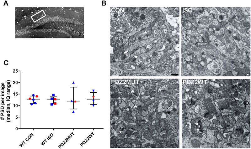
Fig. 4.
Neonatal exposure to ISO or PDZ2WT peptide did not have an acute impact on the number of hippocampal postsynaptic densities. A, dorsal hippocampal region of a semi-thin section illustrating DG subregion of interest (white box). B, representative ultrastructure images from PND21 mice exposed at PND7 to O2 (WT CON, 100% O2), WT ISO (1.5% ISO in O2), PDZ2MUT peptide (8 mg/kg inactive peptide), or PDZ2WT peptide (8 mg/kg active peptide); scale bar represents 500 nm. Asterisks showing some PSDs (not all are labeled). C, plots showing the median with interquartile range number of PSD’s. WT CON (n=6) vs WT ISO (n=5), p=0.829; PDZ2MUT (n=4) vs PDZ2WT (n=4), p=0.742. Data from individual animals are plotted and color coded by gender (red=female and blue=male). Data were analyzed with Mann-Whitney.

