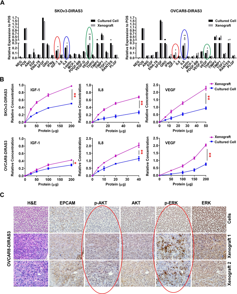
Figure 1. Levels of VEGF, IL-8 and IGF1 are elevated and signaling through PI3K/AKT and MAPK/ERK is activated in ovarian cancer xenografts.
(A) Growth factor/cytokine antibody arrays were performed using cultured cell lysates and xenograft lysates from SKOv3-DIRAS3 and OVCAR8-DIRAS3 ovarian cancer cell lines. Levels of IGF-1 (red circles), IL-8 (blue circles) and VEGF (green circles) were significantly elevated in xenografts when compared with cultured cells (NEG: negative control, POS: positive control). (B) ELISAs were performed to confirm observations in (* p<0.05, ** p<0.01). (A). (C) Phospho-AKT and phospho-ERK levels were increased in xenografts compared with cultured cells. Sections from formalin-fixed, paraffin embedded OVCAR8-DIRAS3 cell pellets and xenografts were examined by immunohistochemistry using antibodies against EpCAM, p-AKT, total AKT, p-ERK and total ERK as indicated.

