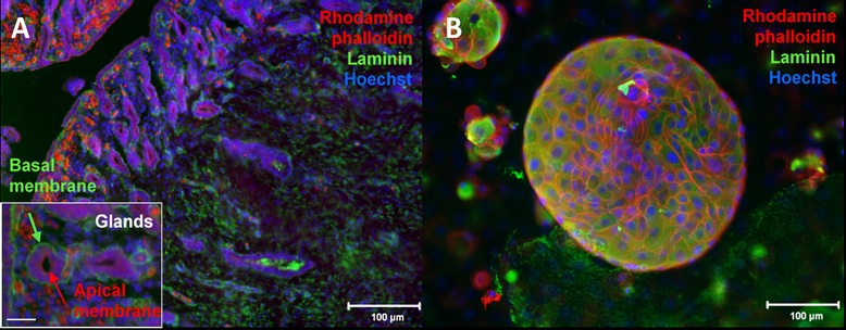
Fig 3. Immunostaining for cell polarity.
Fluorescent micrograph of immunostaining with Rhodamine phalloidin (cytoskeleton = red), laminin (basement membrane = green), and Hoechst (nuclei = blue) (A) 5 μm uterine horn cross-section. Inset: 400X micrograph of two individual glandular structures which show apico-basal polarity where the laminin-rich basement membrane faces away from the lumen. Bar = 50 μm (B) Circular cell structure stained after two weeks of endometrial cell culture.

