
Fig 6. Fluorescent micrographs of 3-D endometrial cell structures after 2 weeks of culture.
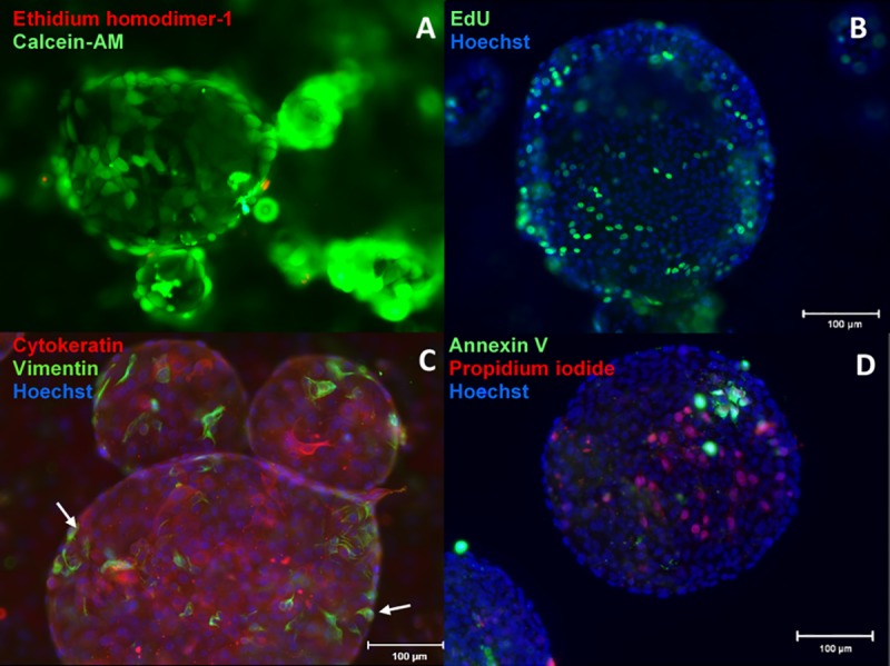
(cell nuclei are counterstained with Hoechst, blue). A) Viability assay with Calcein-AM (live cells) and ethidium homodimer-1 (dead cells) show hollow, lumen-like structures. B) Proliferation assay with EdU showing cells actively synthesizing DNA and proliferating within a spherical cell structure. C) Staining of epithelial (cytokeratin) and stromal cells (vimentin) showing primarily epithelial cells forming the 3-D endometrial cell structures with stromal cells (white arrow) lining the outer surface. D) Detection of early apoptotic nuclei (annexin V) and dead cells (propidium iodide).