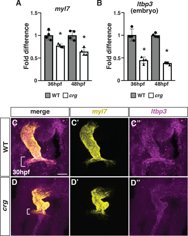
Fig 2. SHF markers of the arterial pole are reduced in crg mutants.
(A,B) RT-qPCR for the pan-cardiac differentiation marker myl7 and SHF marker ltbp3 from embryos at 36 and 48 hpf. (C-D”) Two-color FISH for myl7 and ltbp3 in WT sibling and crg mutant embryos at 30 hpf. Brackets in C and D indicate presence and absence of ltbp3 at the arterial pole of WT and crg mutant hearts, respectively. n = 5 WT and n = 5 crg mutants embryos were examined.

