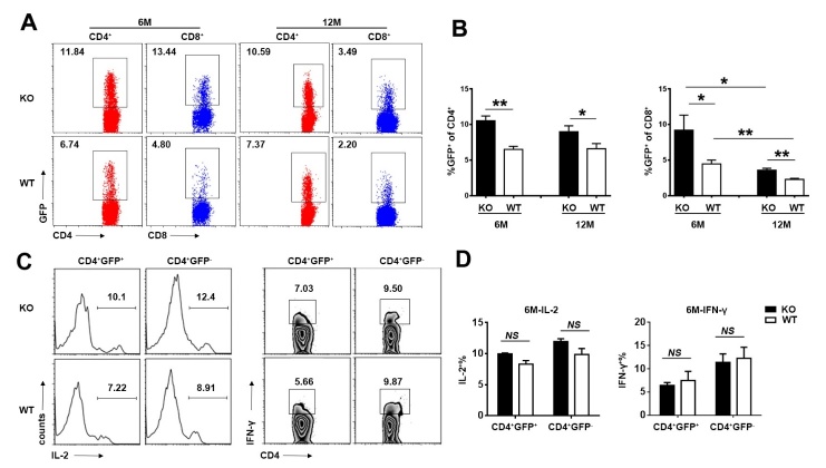
Figure 3. The alteration of recent thymic emigrants (RTEs) in early middle-aged Aire-deficient mice.
(A, B) The proportion of GFP+ cells in peripheral CD4+ and CD8+ cells in 6- and 12-month-old Aire-/- mice and WT littermates. Representative dot plots (A) and the statistical data (B) are shown. (C, D) Splenocytes from 6-month-old Aire-/- and WT littermates were stimulated with anti-CD3 and anti-CD28 for 6h. IL-2 and IFN-γ producing cells in the GFP+CD4+ and GFP-CD4+ T cells was determined by intracellular staining. Representative plots (C) and the percentage of IL-2+ and IFN-γ+ cells (D) are shown. Statistical data are presented as Mean ± SD (n=8 pairs). The experiments were repeated four times. Statistical differences between groups were determined by the Student’s t test. *p< 0.05, **p < 0.01, and NS, no significance.

