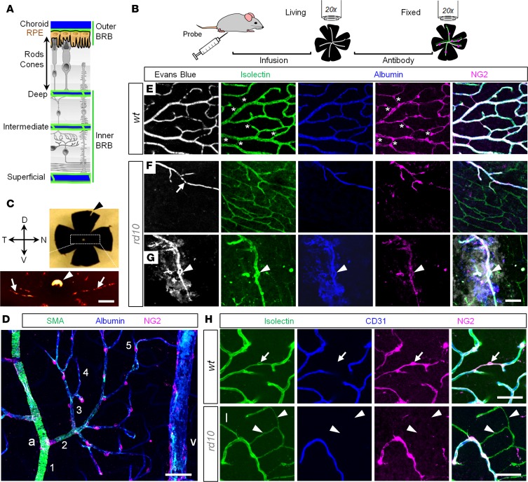
Figure 1. Combinatorial approach for assessment of the vascular network and blood-retina barrier in living and fixed retinal tissue.
(A) A dual nature of blood supply in the retina. Photoreceptors and retinal pigment epithelium (RPE) separate the outer and inner blood-retina barriers (BRBs). (B) BRB assessment protocol. (C) Identification of retinal poles in the eyecup preparation. The nasal-temporal axis is aligned with light choroidal marks (arrows); the dorsal pole corresponds to the white spot of the optic nerve head (arrowhead). (D) Hierarchy of retinal blood vessels. a, artery; v, vein. Numbers indicate branch order. (E–G) Quadruple-labeled confocal sections of the deep vascular layer in WT (E) and rd10 mice at P200 (F and G). A structural marker, isolectin, revealed a net of blood vessels in WT and rd10 retinas (green). In WT retina, all vessels were perfused by Evans Blue (white). In rd10 retina, only a fraction of capillaries was functional (F, arrow). Tortuous blood vessels in rd10 retina were leaky (G, arrowheads). In the fixed tissue, albumin (blue) reproduced Evans Blue pattern. Pericytes, labeled for neuron-glia antigen 2 (NG2), concentrated on perfusable blood vessels (magenta; bridging pericytes are marked by asterisks). In merged images, images were aligned by warp procedure to compensate for fixation-related distortion. (H and I) Isolectin (green) labels basement membrane around pericytes (magenta) and endothelial cells (blue); a bridging pericyte is shown (arrows). Degenerating retinal blood vessels in rd10 retina form empty vascular sleeves without endothelial cells or pericytes (I, arrowheads). Scale bars: 250 μm (C), 50 μm (D–I). SMA, smooth muscle actin; CD31, cluster of differentiation 31.

