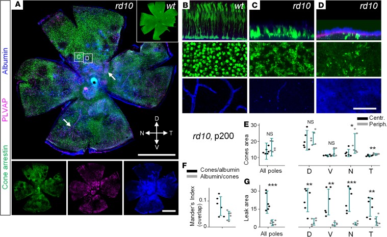
Figure 5. Cone loss and the presence of vascular leak in the advanced stages of retinal degeneration.
(A) In rd10 mice, cones (arrestin, green) were absent in the areas with vascular leak (albumin, blue) coming from fenestrated vasculature (plasmalemma vesicle-associated protein [PLVAP], magenta, arrows). (B–D) Magnified areas from A. In WT, cones were evenly distributed through the retina; no albumin labeling was detected outside of the capillaries. In rd10 mice, regions with leak (D) corresponded to lower densities of cones. Most capillaries of the deep layer were degenerated in rd10 mice. (E) The majority of the surviving cones were found in the periphery, especially of the dorsal pole. (F) Mander’s indices show that areas with surviving cones and areas with blood-retina barrier leak do not overlap. (G) Leak was seen in patches, predominantly across central areas and around deep layer capillaries in the periphery. Scale bar: 1 mm (A); 50 μm (B–D). Data are represented as average ± SD (6 mice, each measurement). Two-tailed t test, *P < 0.05, **P < 0.01, ***P < 0.001.

