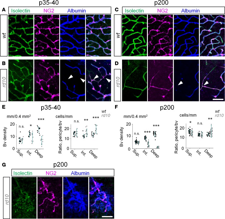
Figure 6. Disruption of blood-retina barrier in RD is not driven by the loss of pericytes.
(A) In WT mice at P35–P40, all capillaries (green) were perfused, as revealed by albumin labeling (blue) and covered by pericytes (magenta). (B) In rd10 mice, degenerating capillaries in the deep and intermediate layers were nonperfusable but contained pericytes (arrowheads). (C and D) At P200, capillaries were well perfused in WT mice while constricted in rd10 mice. (E and F) In rd10 mice, despite vascular decline, the ratio of pericyte to perfused capillaries increased due to persistence of pericyte on degenerated capillaries. (G) Tortuous leaky capillaries in the deep layer in rd10 mice. Scale bar: 50 μm. Average ± SD (4–5 mice). Two-tailed t test, *P < 0.05, **P < 0.01, ***P < 0.001. NG2, neuron-glia antigen 2; bv, blood vessels.

