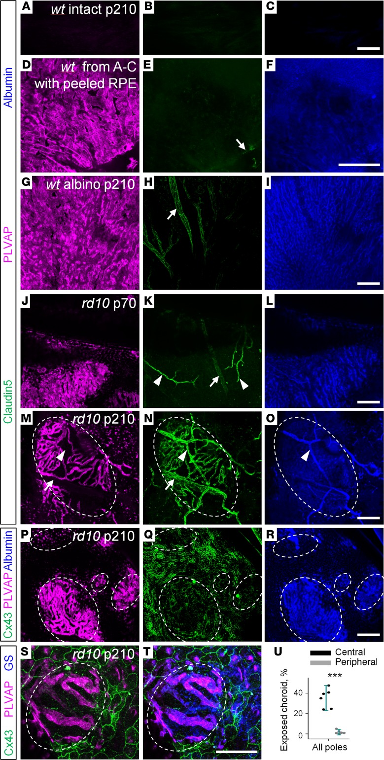
Figure 8. Emergence of blood-retina barrier elements in choroid where retinal pigment epithelium is compromised.
(A–C) In WT eyecup preparation, stained for claudin5, PLVAP, and albumin, no labeling is visible through pigmented retinal pigment epithelium (RPE) cells. (D–F) In pigmented mice from A–C, RPE cells were removed after the eyecup preparation was imaged. (D–I) In WT pigmented and albino mice, choroid capillaries expressed PLVAP (magenta) and did not express claudin5 (green). In contrast, large, sparse, deeply positioned choroid blood vessels were positive for claudin5 and negative for PLVAP (arrow). (J–L) In rd10 mice at P70, exposed capillaries of choroid had no claudin5 expression. Due to the absence of photoreceptors, retinal blood vessels became visible (arrowheads). The arrow shows large choroid blood vessels behind choroid capillary layer. (M–O) At P210 in rd10, choroid gained claudin5 expression in the bare patches without RPE cells (circled). (P–R) In the absence of RPE labeled for Cx43 (green), choroid became exposed and leaky (blue). (S and T) Muller glia, immunostained for glutamine synthetase (GS, blue), were associated with the exposed choroid (outlined by the dashed line). (U) Exposed choroid was mostly present in the central retina. Scale bars: 0.1 μm. Average ± SD (6 mice). Two-tailed t test, ***P < 0.001. PLVAP, plasmalemma vesicle-associated protein; alb, albumin; Cx43, connexin43.

