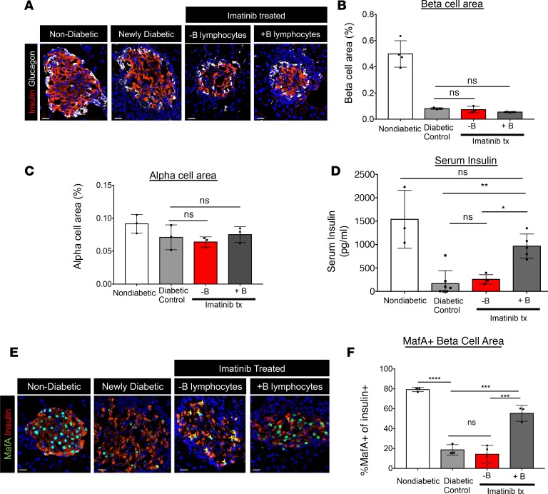
Figure 4. Imatinib therapy restores β cell function but not β cell mass in mice with B lymphocytes.
(A) Pancreatic sections from WT, newly diabetic (2 consecutive blood glucose readings > 200 mg/dl), and imatinib-treated (Imatinib tx) mice with or without B lymphocytes (4 days after treatment initiated) were evaluated for β cell area and α cell area by staining with insulin- or glucagon-specific antibodies. (B and C) Neither β cell nor α cell area increased following imatinib treatment. n = 3 in each group; 2-way ANOVA followed by Šidák’s multiple-comparisons test. (D) Serum insulin levels were partially restored in NOD mice following imatinib treatment. **P = 0.0032, *P = 0.022, 2-way ANOVA followed by Šidák’s multiple-comparisons test. n = 3 control, n = 7 diabetic control, n = 5 imatinib + B lymphocytes, and n = 4 imatinib – B lymphocytes. (E) MafA and insulin staining of pancreatic sections reveals loss of MafA from insulin+ cells in newly diabetic mice. Only imatinib-treated mice with B lymphocytes restored MafA in insulin+ cells. (F) Quantification of MafA+insulin+ cells revealed partial recovery in imatinib-treated mice with B lymphocytes as compared to newly diabetic mice or imatinib-treated mice with no B lymphocytes. (****P < 0.0001; ***P < 0.0005, 2-way ANOVA followed by Šidák’s multiple-comparisons test. n = 3 in each group. Scale bars: 20 μm.

