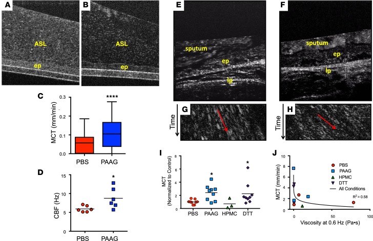
Figure 2. Effect of PAAG on mucus transportability.
(A–D) Representative μOCT images of CF HBE monolayers treated with PBS control (A) or PAAG (250 μg/ml airway surface liquid [ASL] concentration, 24 hours) (B) demonstrated clearly reduced reflectivity of the mucus layer following PAAG treatment, indicating reduced viscosity in situ without altering the integrity of the cell monolayer. ep, epithelium. Quantitative data from μOCT video imaging/rate imaging showed increased mucociliary transport (MCT; C) and improved ciliary beat frequency (CBF; D) following PAAG treatment. n = 6 filters/condition (1 CF donor); *P < 0.05, ****P < 0.0001 by unpaired t test. (E–J) Expectorated CF sputum was treated with PAAG or HPMC (100 μg/ml × 2 hours), or PBS control, and then added to the surface of freshly excised rat trachea while monitoring by μOCT. DTT-treated sputum (100 μg/ml × 2 hours) was added at the end of each experiment as a control. Representative μOCT images from rat trachea after the addition of PBS-treated CF sputum (E) or PAAG-treated CF sputum (F). lp, lamina propria. Time-dependent reprocessed image showing tracks of sputum particles positioned less than 50 μm above the epithelial surface of PBS-treated CF sputum (G) or PAAG-treated CF sputum (H); the more horizontal direction of particle streaks (red arrow) indicates more rapid transport. (I) Summary data showing effect of PAAG on CF sputum transportability. (J) Correlation between MCT and viscosity. n = 3 sputum donors. *P < 0.05 by Kruskal-Wallis test with Dunn’s MCT; r2 = 0.58 for semi-log relationship.

