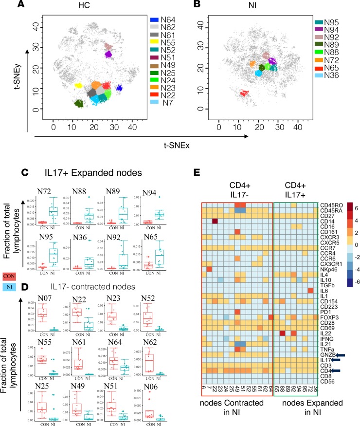
Figure 2. Proinflammatory IL-17–producing CD4+ T cell subsets expand in neuroinflammatory epileptic disease.
(A) The position of CD4+IL-17– nodes on the t-SNE map representing the healthy control (HC) group. CD4+IL-17– nodes were contracted in the neuroinflammatory (NI) epileptic disease group (refractory epilepsy and autoimmune encephalitis) compared with the HC group. (B) The position of proinflammatory CD4+IL-17+ nodes on the t-SNE map representing the NI group. CD4+IL-17+ nodes were expanded in the NI group compared with the HC group. (C) The distribution of each expanded CD4+ node. (D) The distribution of each contracted CD4+ node. (E) The median expression value of surface or intracellular marker for each of the differentially modulated CD4+ nodes.

