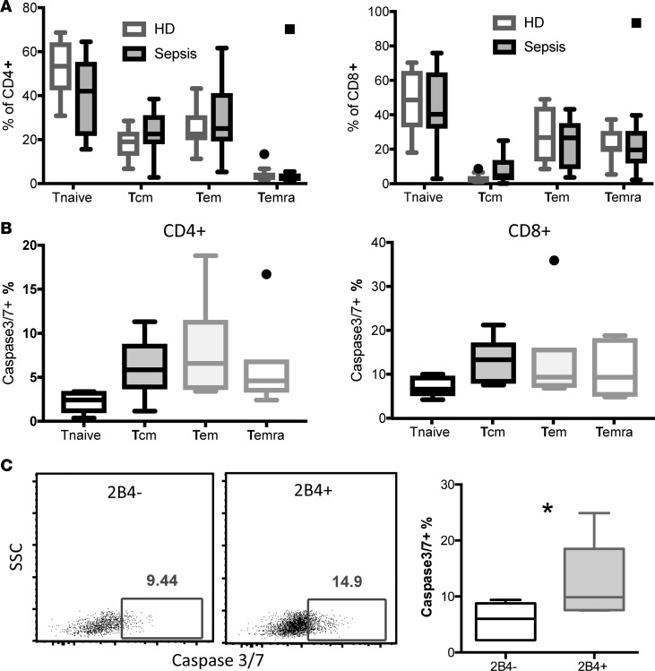
Figure 5. 2B4+CD8+ T cells in patients with sepsis exhibited increased cell apoptosis.
PBMCs were isolated from n = 14 patients with sepsis under an IRB-approved protocol within 24 hours of a sepsis diagnosis and from n = 10 normal healthy controls. (A) Frequencies of memory T cell subsets in n = 10 HDs versus n = 7 patients with sepsis. Naive T cells (Tnaive) were identified by gating on CD45RA+CCR7+; central memory T cells (Tcm) were gated on CD45RA–CCR7+; Tem were gated on CD45RA–CCR7–; and Temra were gated on CD45RA+CCR7–. Cells from septic patients 1–7 as identified in Supplemental Table 1 were used in A. The box plots depict the minimum and maximum values (whiskers), the upper and lower quartiles, and the median. The length of the box represents the interquartile range. (B) Active caspase 3/7 staining on CD4+ and CD8+ T cell subsets isolated from n = 7 patients with sepsis was determined by flow cytometry. Summary data of frequencies of active caspase-3/7+ T cells within memory T cell subsets of CD4+ T cells (left) and CD8+ T cells (right) are displayed. (C) CD8+ T cells were further divided into 2B4– and 2B4+ populations and caspase-3/7 activity was assessed. Representative flow plots for caspase-3/7 staining in CD8+2B4– and CD8+2B4+ T cells. Summary data of frequencies of caspase-3/7+ cells within 2B4–CD8+ and 2B4+CD8+ T cells. Cells from septic patients 8–14 as identified in Supplemental Table 1 were used in B and C. *P < 0.05. SSC, side scatter.

