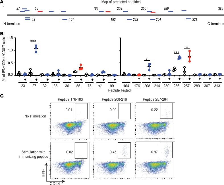
Figure 1. Antigenicity of putative and known epitypic peptides of OVA.
(A) Positions of putative (blue) and the previously reported (red) epitopes of OVA, as detailed in Table 1. Horizontal black line represents the primary structure of OVA. Italicized numbers above and below OVA mark various amino acid positions. (B) Immunogenicity of each putative epitypic (blue) and the previously known (red) peptides of OVA shown in Table 1. “–” Indicates no stimulation and “+” indicates stimulation of LN cells in vitro with the immunizing peptide for 12 hours. Tested peptides are identified on the x axis with the position of their N-terminal residue (n = 3; each peptide tested in 9 different mice over 4 independent experiments; Welch’s t test). (C) Representative assay of the ability of SIINFEKL (257–264), peptide 176–183 (also previously known), and 208–216 (potentially novel peptide) to elicit CD8+ T cell responses upon immunization of C57BL/6J mice, as described in Methods (each peptide tested in 9 different mice in several independent experiments). Flow cytometry plots of viable CD3+CD8+ cells from LNs of immunized mice are shown. *P ≤ 0.05, ***P ≤ 0.001.

