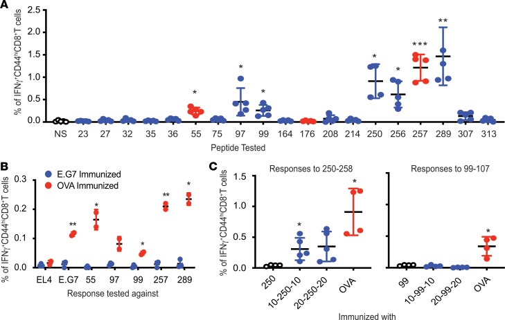
Figure 2. MHC I–restricted epitypicity of OVA.
(A) CD8+ T cell responses to OVA elicited in C57BL/6J mice immunized with OVA emulsified with TiterMax. Responses against each of the putative (blue) and the previously known (red) epitopes of OVA (as listed in Table 1) were tested as in Figure 1. NS indicates no restimulation. P values reflect the significance of difference in percentage IFN-γ–expressing CD44hiCD8+ T cells between NS and peptide-stimulated cultures (n = 5; experiment performed 3 times; Welch’s t test). (B) Inability of irradiated E.G7 cells to elicit CD8+ T cell responses against itself or OVA. CD8+ T cells from LNs of immunized mice were stimulated with splenocytes pulsed with indicated peptide or EL4 or E.G7 cells for 12 hours. P values indicate significance of difference between cultures stimulated with the indicated peptide or cells (n = 3 for E.G7-immunized mice, n = 2 for OVA-immunized mice; experiment performed 4 times; unpaired t test). (C) Extended variants of peptide 250–258 are immunogenic. Mice were immunized with precise peptide (labeled 250 or 99), extended peptides with 10 (10-250-10 or 10-99-10) or 20 (20-250-20 or 20-250-20) flanking amino acids on either termini, or whole OVA. Responses to 250–258 and 99–107 were tested as in Figure 1 (n = 4 for 250 and OVA, n = 5 for 10-250-10 and 20-250-20 in left panel; n = 4 for all sets in the right panel; experiment performed 2 times; Welch’s t test). *P ≤ 0.05; **P ≤ 0.01; ***P ≤ 0.001.

