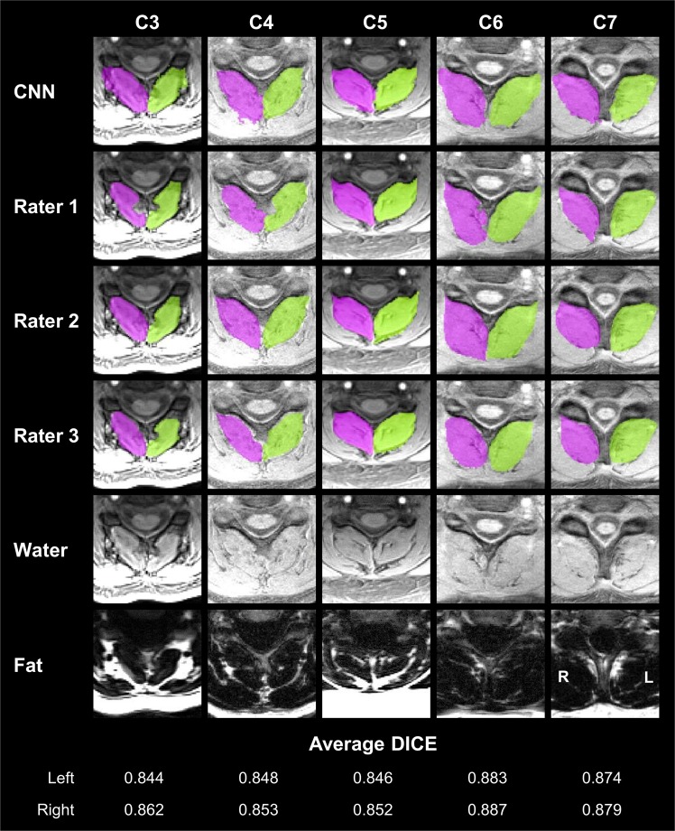
Figure 1.
Convolutional neural network (CNN) segmentation results of the deep cervical extensors. CNN segmentation masks of the left (green) and right (magenta) deep cervical extensors (i.e., multifidus and semispinalis cervicis) are shown from five randomly selected testing datasets. Example axial images at the C3 to C7 vertebral levels were selected to show changes in the deep extensor muscle morphometry across the cervical spine. For comparison, the segmentation masks from each rater are also shown (rows 2–4). The bottom two rows show the water-only and fat-only images for reference. For each example, the average DICE between the CNN and each rater is reported for the left and right masks. The C3 vertebral level is from the inferior portion of the C3 vertebra. L = left, R = right.

