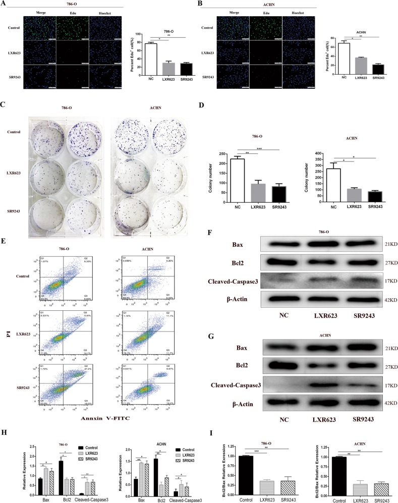
Fig. 2. LXR623 and SR923 inhibit ccRCC cell proliferation and promote apoptosis in vitro.
a, b EdU assay: cell staining using EdU (green) and the nuclear dye Hoechst (blue); 7860-O cells treated with 0.1 µM SR9243 or 1 µm LXR623 for 72 h, ACHN cells treated with 0.1 µM SR9243 or 5 µm LXR623 for 72 h. Scale bar: 100 μm. c, d Colony formation assays: SR9243 (0.1 µM) and LXR623 (0.1 µM and 1 µM) were applied to 786-O and ACHN cells for 72 h. e 786-O and ACHN cells were stained with Annexin-V FITC/PI and analysed via flow cytometry. SR9243 (0.1 µM) and LXR623 (1 µM for 786-O cells, 5 µM for ACHN cells) acted on the two cell lines. f–i Western blotting experiments were performed by treating three groups of cells with DMSO, 0.1 µM SR9243 or LXR623 (786-O, 0.1 µM; ACHN, 1 µM) for 48 h and then extracting proteins. The protein levels of Bax, Bcl2, and Cleaved-Caspase3 were determined via immunoblotting. All data are expressed as the mean ± S.E.M. The experiment was repeated at least three times. Statistical significance was determined using two-tailed Student’s t-test or one-way ANOVA. *p < 0.05; *p < 0.01

