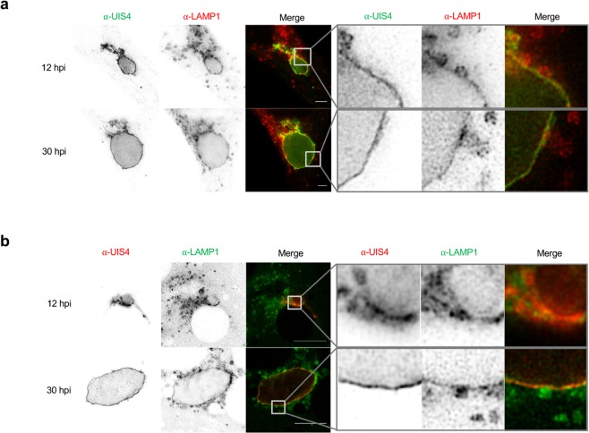
Figure 2.
Super-resolution microscopy reveals that LAMP1-positive vesicles directly associate with the PVM in HeLa cells as well as in primary murine hepatocytes. STED images of HeLa cells (a) and primary murine hepatocytes (b) infected with mCherry-expressing P. berghei (PbmCherry). Images were taken using the Leica TCS SP8 STED microscope. Scale bars are 10 µm. (a) HeLa cells were fixed and stained with α-UIS4 (green; Oregon Green 488) and α-LAMP1 (red; Alexa532). (b) Primary murine hepatocytes were fixed and stained with α-UIS4 (red; Alexa532) and α-LAMP1 (green; Oregon Green 488). Enlargements of the indicated boxed part represented next to each image show possible fusion of vesicles with the PVM. Furthermore, it shows that LAMP1 localizes in the PVM of the parasite. Note that the resolution in the primary murine hepatocytes is decreased compared to the HeLa cells. The cultivation of primary murine hepatocytes requires coating with collagen, which partially interferes with the imaging. Additionally, primary murine hepatocytes are relatively thick compared to HeLa cells, which also decreases the resolution.

