
Figure 1.
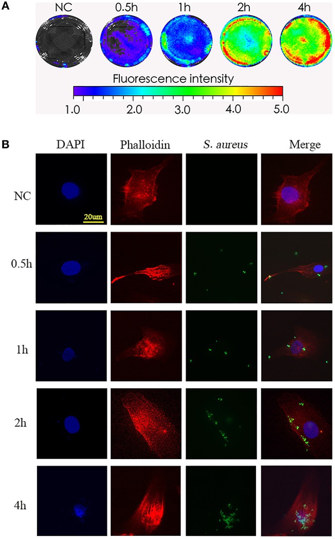
Phagocytosis of S. aureus by NPCs in vitro. (A) NPCs were incubated with bioluminescent S. aureus strains (MOI = 1:10) for different time points (0, 0.5, 1, 2, 4 h), and the bioluminescent intensity was detected using a Xenogen-Caliper IVIS-100 instrument. (B) NPCs were analyzed by confocal microscopy after incubation with GFP-labeled S. aureus for different periods at a MOI = 1:10. The scale bar represents 20 μm. All micrographs are represented at least three experiments.