
Figure 2.
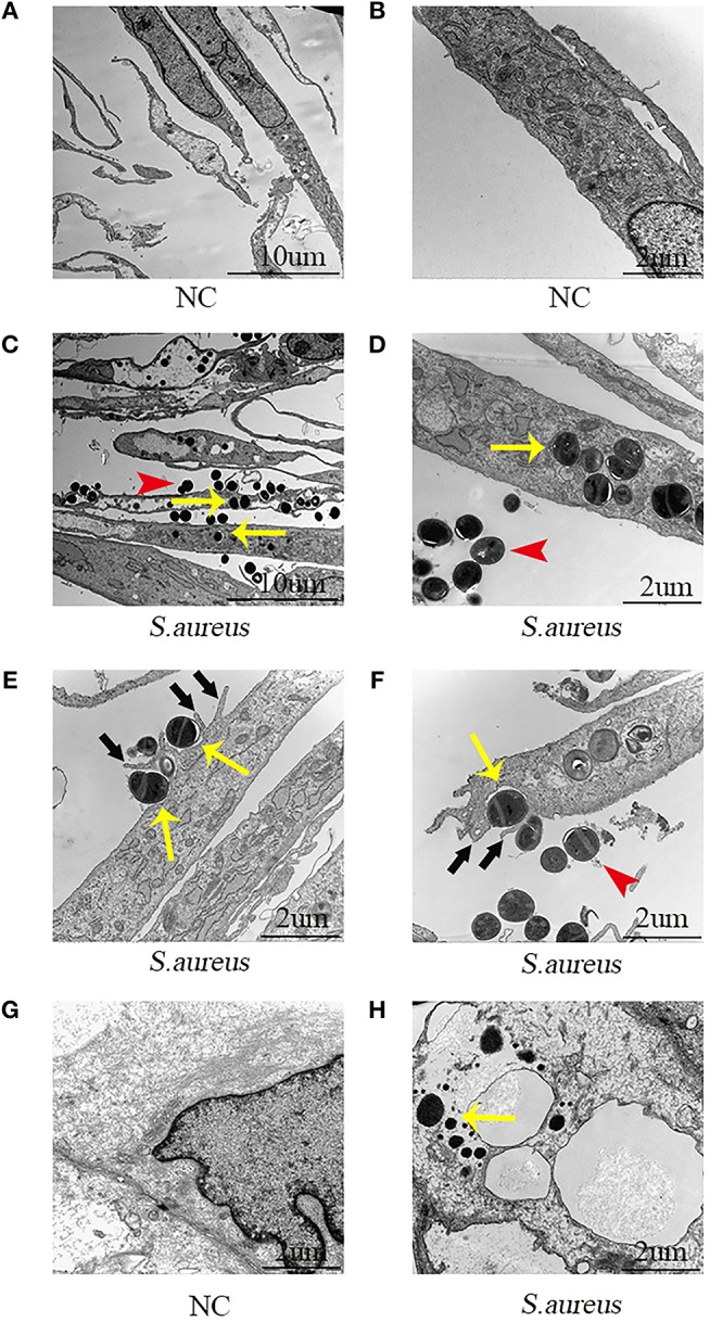
Electron microscopic images of NPCs phagocytizing S. aureus in vivo and in vitro. (A,B) NPCs without incubation with S. aureus were characterized by a large rod nucleus and a thin rim of agranular cytoplasm. (C,D) After co-culturing the NPCs with S. aureus for 4 h, ingested S. aureus (yellow arrow) was observed in the phagosome within the cytoplasm of NPCs and extracellular S. aureus was found (red arrow). (E,F) There were a varying number of membrane protrusions (black arrows) tightly engulfing S. aureus (yellow arrows). (G,H) Compared with NPCs without incubation with S. aureus, ingested S. aureus (yellow arrow) were observed in the cytoplasm of NPCs after caudal IVD infection with S. aureus for 4 h in vivo. All micrographs are represented at least three experiments.